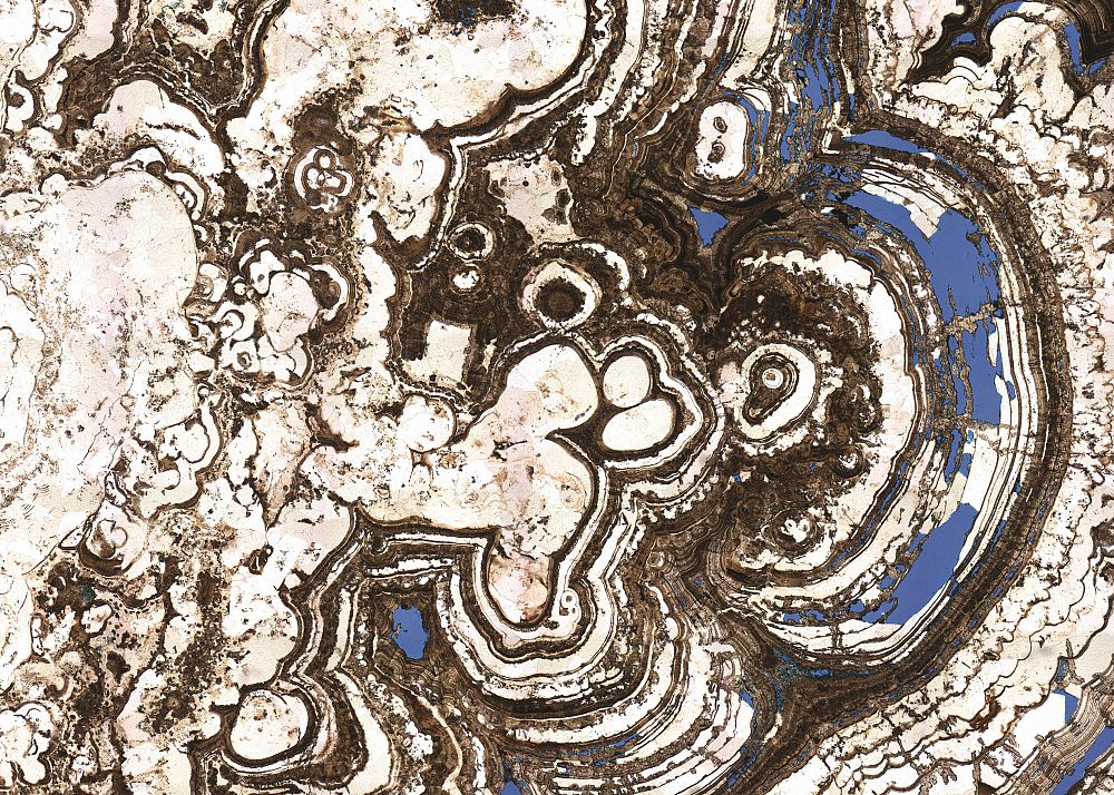
A single image of the entire sample taken at high magnification
We capture a whole thin-section by tiling together a series of photomicrographs; the resulting high resolution image presents an overview of the entire thin section.
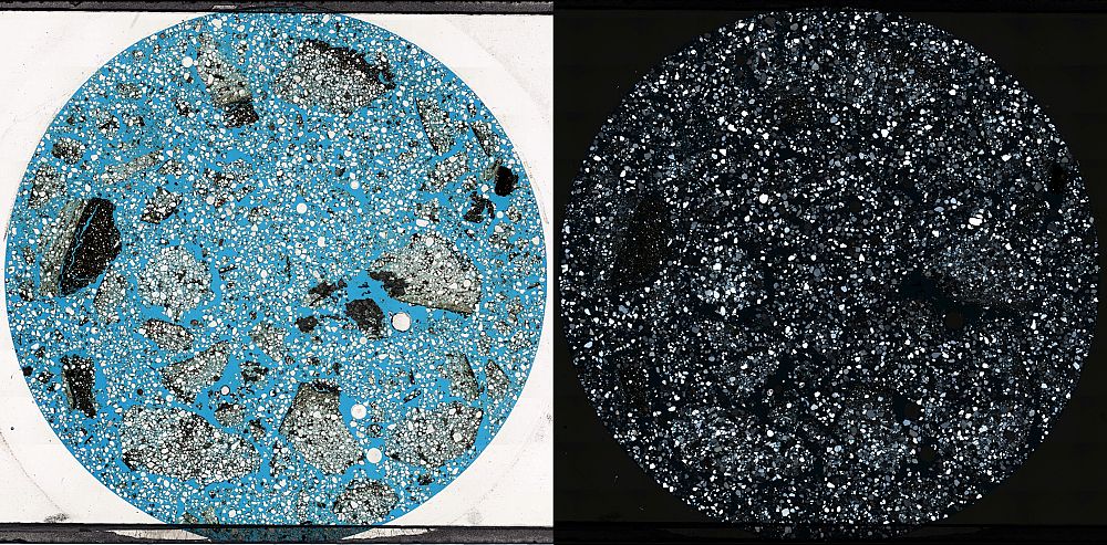
High-resolution imaging of whole thin-sections in plane-polarised and cross-polarised light creates a permanent and easily accessible digital archive for collaboration purposes and image analysis techniques
A single image of the whole sample with the ability to zoom in without pixilation.
Finely calibrated to allow for micron-precision measurements, away from the microscope.
Image output as uncompressed TIF and compressed JPG format, compatible with any image software.
Ability to conduct Petrographic Image Analysis (PIA) techniques to provide detailed additional data on your thin-sections.
Fully automated capture process
The capture process is fully automated; the thin-section is typically run twice, with plane-polarised and cross-polarised light.
The compiled images can be saved in a variety of formats – uncompressed TIF being the highest quality.
The software employs an overlap technique to capture the full image.
Finely calibrated pixel-to-micron conversion data to allow the user to accurately measure objects on screen, eg. grain size and pore space.